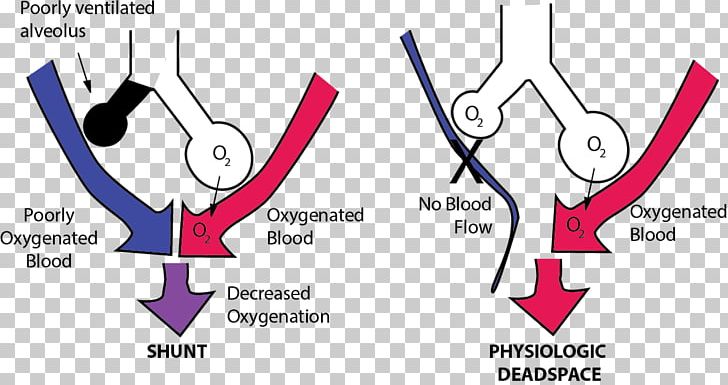

If a substantial proportion of lung units have a very high V/Q (e.g.

Normally, there is only mild heterogeneity of V/Q ratios but this is increased in various pathological states. This is the most difficult to grasp but also perhaps the most important factor in many disease states.

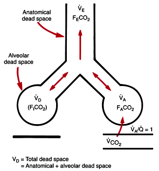
A V/Q of infinity is the definition of alveolar dead space. Since the perfusion of the lung unit reduces to zero, the V/Q ratio for this unit is infinity (V÷0 = ∞). Consider a scenario where a small embolus blocks all perfusion to a lung unit. Normally, ventilation and perfusion of respiratory zones (lung units) are nicely matched (normal V/Q ~0.9). The dead space in an average adult has been reported to be ~150 cc or 2cc/kg ideal body weight. Anatomical dead space is thus defined as the volume of the conducting zone (Figure 1). The remaining circuit: respiratory bronchioles to alveolar sacs (generation 23) participate in gas exchange and is called the respiratory zone. The entire airway circuitry all the way from mouth to the terminal bronchioles (~generation 14-16) is the conducting zone of the respiratory system. Mechanisms that create ‘dead space effect’.A conceptual, simplistic categorization is as follows: The nomenclature describing dead space can be quite confusing (see for in-depth reading). , where V̇CO 2 = CO 2 production by the body (units: cc/min) and V̇ A = alveolar ventilation. The alveolar ventilation controls CO 2 homeostasis according to the alveolar ventilation equation : Mathematically, V̇ A is total ventilation minus anatomical dead space ventilation. Its calculation requires exclusion of ventilation occurring in the anatomical dead space. in a passive mechanically ventilated patient on volume control (VC) mode, V̇ E = V T x RR.Īlveolar ventilation ( V̇ A the subscript ‘A’ denotes ‘alveolar’) is the amount of ventilation occurring in the alveoli in one minute. Minute ventilation ( V̇ E the subscript ‘E’ denotes ‘exhaled’) is the total ventilation in one minute (units: L/min). At this point, it is helpful to define minute ventilation and alveolar ventilation : V D /V T is a common way to quantify dead space. The fraction of the tidal volume that does not contribute to gas exchange is known as dead space fraction (V D /V T where V T = tidal volume and V D = dead space volume). A certain amount of dead space is normally present in every person (this is known as anatomical dead space: see below).

This concept can be extended to include factors that cause a dead space effect. Simply put, dead space represents the volume of ventilated air that does not participate in gas exchange.


 0 kommentar(er)
